3D Printed Organs: Innovations in Bioprinting Technology
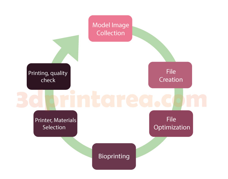
What are 3D Bioprinting and Biofabrication?
Tissue engineering—encompassing the development of 3D printed organs and other tissue structures—aims to repair damaged tissues or organs. However, traditional tissue engineering methods have struggled to achieve clinical success in processes requiring complex production or repair due to certain limitations. At this point, biofabrication offers a significant solution. In today’s rapidly advancing technological landscape, 3D bioprinting, a part of biofabrication and an extension of additive manufacturing, has begun to be utilized in applications where traditional tissue engineering falls short.
Advantages and Disadvantages of Bioprinting
Advantages of Bioprinting
- Precision Medicine: From the initial to the final stages, bioprinting allows the creation of patient-specific structures using their unique anatomical data through CAD software. This reduces the risk of incompatibility between the final product and the patient.
- Advanced Biomaterials and Cell Innovation: Advanced biomaterials provide biocompatible and biodegradable properties that can integrate with the patient’s tissues over time. These innovative materials, with their adaptability and compatibility, make 3D bioprinting reliable and preferable.
- Applicability to Different Tissue Types: Beyond skin tissue, the processes can be adapted for the production of muscle, cartilage, and bone tissues.
- Rapid Prototyping: One of the greatest shared benefits of 3D printing technologies across all application areas is rapid prototyping. In 3D bioprinting, productions that would typically take a long time with traditional methods can be completed in much shorter periods. This capability can be life-saving in emergencies.
Disadvantages of Bioprinting
- Complex structures and vascular networks: During the production process, the creation of vascular networks is essential to ensure the tissues remain viable. In the production of complex organs, generating these networks in a detailed and functional manner presents certain technical challenges.
- Diverse cell types: Organs and tissues are not composed of a single type of cell. During production, these cells must be arranged accurately, which involves a technologically complex process.
- Cost: These processes are costly due to factors such as the need for advanced technological equipment, the high cost of biomaterials, and the large budgets required for R&D expenses. Additionally, the legal aspects of working with human organs or tissues may lead to additional costs.
- Integration process: Long-term stability issues may arise during the integration of the printed tissue or organ into the patient’s body.
Let’s Review the Study “3D Printed Organs: The Future of Regenerative Medicine” Published in the Journal of Clinical and Diagnostic Research
The study discusses practical 3D bioprinting techniques as well as clinical application areas.
Key 3D Bioprinting Methods
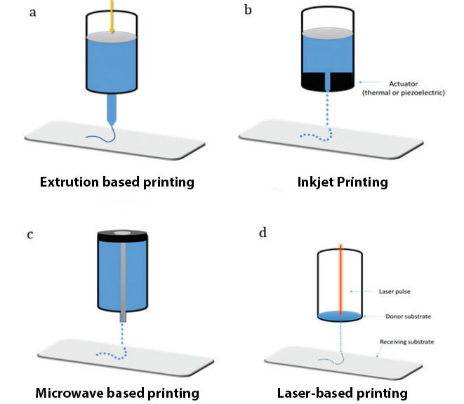
The primary types of printing used in the bioprinting sector are listed as Extrusion Bioprinting, Inkjet Bioprinting, and Stereolithography
- Extrusion Bioprinting: Known for its ability to handle high cell densities and perform well in complex processes, this technique creates 3D structures by controlled extrusion of bioink, which contains biomaterials and cells, layer by layer through a print head.
- Inkjet Bioprinting: This method forms 3D structures by ejecting bioink as tiny droplets through a nozzle. Thermal and piezoelectric techniques are the most commonly used methods. It is a fast and cost-effective method.
- Stereolithography: In this technique, photosensitive polymers are solidified layer by layer using ultraviolet (UV) light. Its advantages include high production precision and the ability to create complex structures.
3D Bioprinting and Biofabrication Application Areas
Blood Vessels:
A research group has successfully developed vascularized tissues composed of various cell structures. These tissues were reported to maintain their cell viability and structural integrity under in vitro conditions for 48 hours.
Another group has produced small-diameter vascular structures using the rapid prototyping bioprinting method. Similarly, another study utilized hydrogels based on sodium alginate and polyethylene glycol diacrylate (PEGDA) to create aortic valves.Liver:
Liver bioprinting has been used in the treatment of liver fibrosis and other complex liver disorders. A group of researchers performed liver bioprinting by combining HepaRG cells with human stellate cells using the stereolithography 3D printing technique. The bioprinted organ showed stable metabolic indicators, proving its reliability for applications such as drug testing and disease modeling.Cartilage:
Cartilage, which lacks blood and nerves and has a unique structure, is known to be challenging to treat. Traditional methods cannot fully heal cartilage tissues. However, 3D bioprinting appears to overcome these difficulties. Researchers successfully bioprinted human joint cartilage using bioink, demonstrating that cartilage defects can be repaired through 3D printing.Muscle:
Musculoskeletal injuries can reduce individuals’ quality of life and impose certain limitations on their daily routines. Once again, 3D bioprinting technology provides innovative solutions for repairing these damages. In previous years, an integrated muscle-tendon unit containing four different components was successfully printed.Bone:
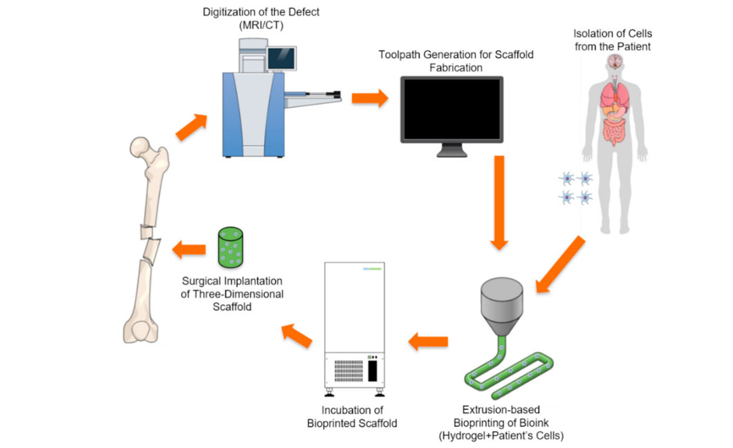
The primary causes of bone injuries include aging, infections, and tumors. Common methods for bone repair, such as allografts or xenografts, come with limitations, requirements, and risks. 3D bone printing technology offers promising solutions to overcome these disadvantages.
The success of bone bioprinting relies on the availability of suitable biomaterials and cell types. In one study, solid bone formation was achieved using PEGDMA hydrogel and biologically active peptides.Skin:
Traditional skin substitution methods are not suitable for personalized treatments and incur additional costs due to frequent replacements during treatment. However, laser-assisted 3D bioprinting can create multicellular 3D structures.
Recently, functional 3D skin bioprinting has been achieved with the help of some biomaterials. Ensuring new blood vessel formation is critical for the success of skin implants. Research has shown that when cells are printed onto hydrogels via 3D bioprinting, cell viability rates remain around 90%. Additionally, scientists have used different cells together to support the skin structure, thus accelerating the healing process.
Recent Developments
An intriguing study published in the journal highlights the restoration of natural reproductive abilities in mice through the use of a 3D-printed microporous bioprosthetic ovary. This bioprosthetic ovary, implanted in mice, promoted vascularization, enabling the mice to give birth. It was reported that the offspring born naturally were raised on their mother’s milk.
Another advancement in this field involves the treatment of meniscus tears using 3D printing technology. Meniscus tears, which occur in the complex cartilage structure of the knee joint, can cause serious problems. The journal mentions a study where sheep menisci were replaced with 3D-printed scaffolds, leading to meniscus regeneration. The study, monitored over a three-month follow-up period, demonstrated that the regeneration of complex tissues is indeed possible.
References
Badwaik, R. (2019). 3D printed organs: The future of regenerative medicine. Journal of Clinical and Diagnostic Research, 13(11), FE01–FE08.
Ramesh, S., Harrysson, O. L. A., Rao, P. K., Tamayol, A., Cormier, D. R., Zhang, Y., & Rivero, I. V. (2021). Extrusion bioprinting: Recent progress, challenges, and future opportunities. Bioprinting, 21, e00116. https://doi.org/10.1016/j.bprint.2020.e00116